Costochondritis (Tietze’s syndrome)

I’ve been asked –and consulted- about “rare chest pain” in several times. I could notice that people is always worried about any kind of chest pain because the fear of suffer a heart attack or a pulmonary problem. Chest pain is one of the most common symptoms that require medical attention. You –as physician- should always exclude this topics (cardiac and pulmonary) in the first place. You should keep in mind that there is a disease called costochondritis (Tietze’s syndrome) once you ruled out the main fear conditions (pulmonary and cardiac).

Costochondritis
is an inflammation of the costo-sternal joint (rib-sternum) or it could be an inflammation between the costo-chondral joint (rib-rib cartilage). The group mainly affected is that woman over 40s.

Etiology (causes)
: Direct injury to the chest, viral infections (cold / flu), idiopathic (the cause cannot be found).

Its clinical manifestations: Pain, tenderness in those joints I already mention earlier. This pain and/or tenderness get worse when you touch the involved site or move in a certain direction.
The diagnosis it’s mainly a clinical one and the physician should always exclude a heart attack and other important things.

The gold-standard of treatment is NSAIDs (non-steroid anti-inflammatory drugs like aspirin, diclofenac, naproxen, ibuprofen, acetaminophen, etc.) for one or two weeks (this disease usually lasts for this period of time). Some patients respond well to putting a local heating pad.

You have to remember that when you have chest pain, you have to look for a health care provider immediately to exclude other serious conditions.

Regards,

Jon Mikel Iñarritu, M.D.

Spanish (Español) article

Insulinoma & Whipple triad – Surgery (update)

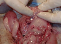
Insulinoma

Insulinoma is the most common functioning tumor of the pancreas, and affected patients present a tableau of symptoms referable to hypoglycemia (symptoms of catecholamine release), mental confusion and obtundation. Many patients have symptoms for years. Some have been greatly troubled by emotional instability and fits of rage, often followed by somnolence.

Incidence: This tumors are so rare. Probably the best series of insulinomas is from the Olmsted County (Mayo Clinic). This study (cohort) was seen over a sixth decade period (’27 to ’86). The incidence there was 0.4 per 100,000 person-years (or four cases per million per year).

The diagnostic hallmark of the syndrome is the so-called Whipple triad, wich consists in symptoms of hypoglycemia (catecholamine release), low blood glucose level (40 to 50 mg/dL), and relief of symptoms after intravenous administration of glucose. The triad is not entirely diagnostic, because it may be emulated by factitious administration of hypoglycemic agents, by rare soft tissue tumors, or occasionally by reactive hypoglycemia. The clinical syndrome of hyperinsulinism may follow one of two patterns or sometimes a combination of both.

The symptom complex may be due to autonomic nervous overactivity, expressed by fatigue, weakness, fearfulness, hunger, tremor, sweating, and tachycardia, or alternatively, a central nervous system disturbance with apathy (or irritability or anxiety), confusion, excitement, loss of orientation, blurring of vision, delirium, stupor, coma, or convulsions.

Because cerebral tissue metabolize only glucose, prolonged profound hypoglycemia may cause permanent brain damage. Clinicians need to be alert to this when attempting to induce hypoglycemia by fasting.
Preoperative NPO orders must be accompanied by intravenous administration of glucose.

The diagnosis: Is established by demonstrating inappropriately high serum insulin concentrations during a spontaneous or induced episode of hypoglycemia. Then imaging techniques are used to localize the tumor. The available procedures include spiral CT, arteriography, ultrasonography (transabdominal and endoscopic), and 111-In-pentetreotide imaging. Transabdominal ultrasonography is our preferred initial test.

Insulinomas are small (usually smaller than 1.5 cm), usually single (only 10% are multiple and those are usually associated with MEN 1 syndrome), usually benign (only 5% to 10% are malignant), and usually hard to find. Success in localization often parallels the degree of invasiveness of the study.

Its treatment:
Treatment for insulinoma is surgical. At operation, the incision is dictated by operator preference, either a midline incision from the xiphoid to below the umbilicus or a bilateral subcostal incision. Exposure should be generous, and mechanical ring retractors are an asset. The entire abdomen should be explored, with particular attention being paid to possible liver metastases.

The head of the pancreas should be palpated carefully and examined anteriorly and posteriorly; the body and tail of the pancreas should be palpated, dividing any ligamentous attachments to the spleen, delivering the spleen into the wound, and rotating the tail anteriorly to allow palpation and visualization.

Anyone operating patients with pancreatic islet adenomas should be familiar with techniques for, and limitations of, intraoperative ultrasonography. The higher-resolution (7.5-to 10-MHz) transducers are used in the pancreas; because of its greater depth of penetration. Islet tumors are detected as sonolucent masses, usually of uniform consistency. Several reports attest to the high degree of accuracy of intraoperative ultrasound.

Review of current literature leads to the almost startling conclusion that nearly all syndromes of hyperinsulinism are due to insulinomas and that nearly all insulinomas can now be detected before or during surgery. Having said this, there appears to be scant justification for an empirical (“blind”) partial pancreatectomy except in patients with insulinoma plus MEN 1.

Most insulinomas are benign and can be enucleated. Nutrient vessels in the bed of the adenoma should be cauterized.

The 10% of patients with hyperinsulinism who have MEN 1 syndrome have multiple islet tumors, one of which is usually dominant and responsible for the excessive insulin output.
Insulinoma

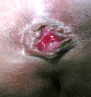
Thrombosed Hemorrhoid

Thrombosed Hemorrhoid

Thrombosed external hemorrhoids often occurs in grade III-IV internal hemorrhoids, and can cause excruciating pain, and patients will often present acutely. In such cases, surgical evacuation of the hemorrhoid can produce immediate relief.
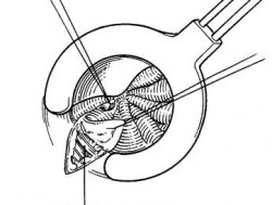
The treatment of choice is hemorrhoidectomy with partial lateral internal sphincterotomy, which consists in incised the mucosa on each side of the hemorrhoid and extended outward toward the anoderm. The anoderm and the hemorrhoidal mass are elevated off the transverse fibers of the internal sphincter muscle. Then the mucosa is approximated with continuous 3-0 absorbable sutures, leaving 2 to 3 mm of the anoderm left open for drainage.

hemorrhoidectomy

Regards,

Jon Mikel Iñarritu, M.D.

Link to: Hemorrhoidal Prolapse

A rare thoracic tumor

Useful links:

50 y/o male with a history of one year with respiratory symptoms, he was diagnosed treated at Guatemala where he lives. Now he comes to Mexico City seeking for a second opinion and other options of therapy. At the physical examination he is well, has diminished breath sounds and just sinus tachycardia.

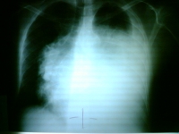
Here is his chest x ray:
Chest x-ray

What’s the diagnosis?

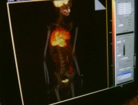
A PET-CT was done:
PET-CT

What’s your diagnosis?

Thymic Carcinoma

Thymic carcinoma displays distinct morphology and biology, different in many ways from thymoma. It is composed of highly atypical cells with cytoarchitectural features of carcinoma similar to those seen in other organs. Although many lymphocytes can be seen in its stroma, they are of B cell type and mature T cell type; thymic carcinoma lacks the immature T cell lymphocytes of thymoma.
Thymic carcinoma is classically not associated with paraneoplastic syndromes such as myasthenia gravis. However, one study reported 22 cases of primary thymic epithelial neoplasms which showed combined features of thymoma and thymic carcinoma; the carcinoma and thymoma were either found synchronously, or carcinoma developed with a preexisting thymoma after 10 to 14 years.
Incidence Primary thymic carcinoma is a rare neoplasm. At the National Cancer Center of Tokyo, 17 cases were resected over a 29 year period, during which time 79 thymomas and 2500 lung cancers were treated.
Clinical and radiographic manifestations Primary thymic carcinoma is a tumor of adulthood (mean age of 46 to 54 years). Most patients complain of chest symptoms, including pain, cough, superior vena cava syndrome, or general symptoms such as fatigue, weight loss, anorexia; fewer than 10% of patients are asymptomatic. Chest radiographs and computed tomography show an anterior mediastinal mass infiltrating along the pleura or mediastinum, with frequent necrosis or rare calcifications.

technorati tags: , , , , , , , , , , , , , ,

New AACE guideline to achieve glycemic goals

Via AACE from J Davidson, P Jellinger, L Blonde, H Lebovitz, C Parkin (2005):

The new position statement is this: Even if the initial A1C is from 6 to 7%, you should start pharmacotherapy.

This aggressive treatment has proved that mortality diminishes considerably.

Regards,

Jon Mikel Iñarritu, M.D.

technorati tags: , , , , , , , ,

Open Fractures Classification and its clinical manifestations

Open fractures are surgical emergencies because of its complications (soft tissue damage, infection, hemorrhage).

We at emergency department need a classification system that it is intented to determine the degree of that damage.

We have to make a rapid (also a good) interrogation of lession mechanisms (what, when,  where, who, associated lessions). REMEMBER: If a low-grade open fracture has 6hrs from the begining, it’s considered infected.

On 1984, Gustilo and Anderson made a classification system, wich is the following:

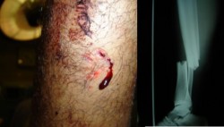
Type I open fracture. Wound less than 1 cm, without contamination and minimal injury of soft tissue.
Type I open fracture - 1
Type I open fracture - 2


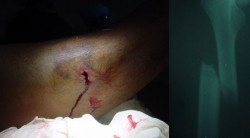
Type II open fracture. Wound between 1 and 10 cm, mild contamination, extensive soft tissue damage and minimal to moderate crushing component.
Type II open fracture - 1
Type II open fracture - 2

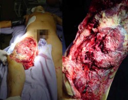
Type III-A open fracture. Wound larger than 10 cm, severe contamination and severe crushing component.

Type IIIA open fracture

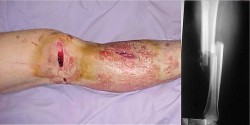
Type III-B open fracture. Wound larger than 10 cm, severe contamination and severe loss of tissues.
Type IIIB open fracture - 1
Type IIIB open fracture - 2

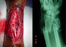
Type III-C open fracture. Wound larger than 10 cm, severe contamination and neurovascular injury.
Type IIIC open fracture - 1
Type IIIC open fracture - 2

REFERENCES:
Gustilo, R.; Mendoza, R.; Williams, D. Problemsin the management of type III (severe) open fractures: A new classification of typeIII open fractures. J Trauma 24:742, 1984.

Regards,
Jon Mikel Iñarritu, M.D.

technorati tags: , , , , , , , , , , , , ,

Cardiac Tamponade

Physical findings & Clinical Presentation:

• Severeconstant pleuritic pain that localizes over the anterior chest and may radiateto arms and back; it is differentiated from myocardial ischemia, becausethe pain intensifies with inspiration and is relieved by sittingup and leaning forward.
• Pericardial friction rub is best heard with patientupright and leaningforward and by pressing the stethoscope firmly against the chest; itconsists of three short, scratchy sounds:
• Signs and symptoms:

  1. Dyspnea
  2. Orthopnea
  3. Interscapular pain
  4. Tachycardia
  5. Refractary hypotension
  6. Distended neck veins
  7. Paradoxicalpulse
  8. Chest x-ray: Cardiomegaly, clear lungs (may be normal)
  9. ECG: Low amplitude of QRS, electrical alternans

Treatment:
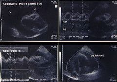
Immediatepericardiocentesispreferably by needle paracentesis with the use of echocardiography, fluoroscopy,orCT Aspirated fluidshould be sent for analysis (protein, LDH, cytology, CBC, Gram stain, AFBstain) and cultures for AFB, fungi, and bacterial C&S.

•

tamponade

Cardiac Tamponade

tamponade

Cardiac Tamponade

Regards,
Jon Mikel Iñarritu, M.D.

technorati tags: , , , , , , , , , ,

Horrible: Child abuse

Another case of Child abuse was shown today on TV news.

It seems that we can’t stop this and some parents preffer to remain in silence rather than to speak out loud about this impunity.

I think it is time to recognize this cases and report them to the authorities. So today’s matter is to know the facts about child abuse & neglect.

Also know as child maltreatment it is a group of comission, omission and lack of actions, that result in morbidiy and/or mortality.

It is every agression or omission of intentional nature, indoor or outdoor, against child(s), before or after the birth that affects their bio-psycho-social integrity. This actions can be from an individual, a group of people (society), an institution taking advantage of their physical and/or intelectual superiority.

There is a triad of child abuse:

  1. Child: Often a non-accepted (desired) kid, malformation, chronic or acute diseases.
  2. Agressor: Often a parent or caretaker, alcoholic or drug abuse problem, background of abuse.
  3. Triggers: Unemployment, marital disfunction, social and economic problems.

It’s importance: 2,900,000 cases / year on U.S.A of wich 33% is physical abuse. Second leading cause of death in child less than 5 years old.

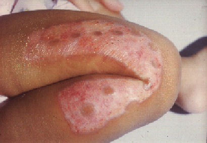
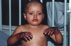
Burn by iron Circular burn

Diagnosis:

You should always suspect child abuse when:

Clinical aspects on child: Non-correlated history given by parents, small height, poor hygiene, previous lesions, malnutrition, non-desired/non-accepted child, incomplete vaccination schedule.

Clinical aspects on child’s family: Delayed medical attention, abuse background, step-mother/father, alcoholism, ask for medical attencion by someone else outside the central group of family.

It is required a multidisciplinary team: Primary care Physician, Pediatrician, Social work, Mental Health, Lawyer, Voluntaries.

The action of preventive mesures should include all levels of society: International organizations, Federal instances, Local community (church, school, friends, pediatrician, etc), Child’s family and Child.

Who where ever you are do: Let’s stop this.

Regards,

Jon Mikel Iñarritu, M.D.

technorati tags: , , , , ,

Concern about appearence

24-year-old male comes to you with the chief complaint that his nose is too big to the point of being hideous. He has been avoiding contact with others and has left his studies.

The patient states that his nose is a constant embarrassment to him and he would like it surgically reduced. He tells the physician that three previous plastic surgeons had all refused to operate on him because they said his nose was fine, but the patient states that “they just didn’t want such a difficult case”. You observe that the patient’s nose is of normal size and shape.

What do you think?

This is a typical manifestation of body dysmorphic disorder (also know as dysmorphophobia), that is an extreme feeling of dislike or concern about some aspect of the appearence in spite of a normal or nearly normal shape The fear of being ugly or repulsive is not decreased by reassurance and compliments and has almost a delusional quality. The social, academic, and occupational lives of this individuals are greatly affected, due to avoidance of social interactions for fear of embarrassment, the time spent in checking mirrors and seeking surgical treatment or cosmetic remedies, and the chronic emotional distress that accompanies the disorder.

Complications: As the presentation of this patient, social withdrawal is common, and patients may be unable to work or to sustain relationships with others. Suicidal ideation is common, and completed suicide may occur.

Treatment: As this patients are loath to consider their pathologic concerns per se, they rarely stay in treatment. Both fluoxetine and clomipramine are effective in reducing the intensity of the patients’ concerns, and there is some preliminary evidence for the effectiveness of behavior therapy.

So, what you don’t like about yourself?

Regards,

Jon Mikel Iñarritu, M.D.

technorati tags: body dysmorphic disorder, dysmorphophobia, unbounded medicine, psychiatry, medicine