
Author: AnaSerrano
What is the diagnosis?
50 y/o woman history of respiratory simptoms.


Pemphigus and bullous pemphigoid. 70 y/o woman with cutaneous lesions
This woman has this cutaneous lesions distributed all over the body. She has history of vitiligo.

Pemphigus and bullous pemphigoid.
These are autoimmune blistering diseases. They are caracterized by the production of antibodies directed against different epitopes, resulting in intraepidermal blistering in pemphigus and subepidermal blistering in pemphigoid. The term pemphigus comes from the greek, pemphix, that means bubble. It was used to describe most blistering diseases until 20th century.
Pemphigus
Pemphigus is a group of rare, chronic, autoimmune and potentially fatal diseases of mucous membranes and skin.Three types of pemphigus have been described:
Pemphigus vulgaris
Pemphigus foliaceus
Paraneoplastic pemphigus
Is caracterized by the production of autoantibodies, in pemphigus vulgaris IgG, against adhesion proteins. This alters the intercellular cement that holds epidermal cells together. Resulting in blister formation. Clinically is caracterized by flaccid bullae that tipically begin in the oropharynx and then may spread to involve the skin. With a predilection for the scalp, face, chest, axillae and groin.
Bullous Pemphigoid
Mostly afects persons 60 yeard of age and older. Pemphigoid refers to pemphigus-like blistering disease but is less aggressive than pemphigus vulgaris. It is also a chronic and recurrent disease of autoimmune origin. As in pemphigus vulgaris, the precise reason for the formation of autoantibodies is unkown. A small group of patient bullous pemphigoid is drug-induced. Penicillamine and furosemide are the most frequently implicated. A common presentation is a widespread blistering eruption in a middle age or eldery person who is taking multiple medication.
The case above was diagnosed as bullous pemphigoid.
Regards,
Gerardo Morales-Mora, M.D.
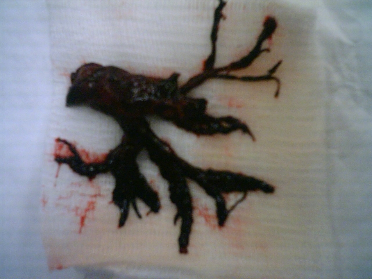
What is this?
A friend from the “INER” National Institute for respiratory diseases, show me this image. It was taken after a extraction from bronchoscopy in a man with atelectasis of the lung.
Regards,
Gerardo Morales, M.D.
Surgical treatment for ingrowing toenail
The patient is a young man with onychocryptosis secondary to overweight. At the images below a matricectomy with remotion of soft tissue are shown. This procedure is performed bedside in few minutes and only need local anesthesia. The goal is to release the nail from the surrounding tissue.


Gerardo Morales, M.D.
CT Guided percutaneous needle biopsy of pulmonary masses
CT
Percutaneous apiration of a pulmonary tumor can be performed through the chest wall, using CT scanning to guide the placement of the needle within the lesion. The yield froma a percutaneous approach is generally higer than the yield from fiberoptic bronchoscopy. Overall, the diagnostic yield from percutaneous needle aspiration has been reported to range from 43 to 97 percent

Gerardo Morales, M.D.
technorati tags: CT, pulmonary mass, biopsy, percutaneous, medicine, unbounded medicine, unboundedmedicine
What if an earthquake destroys Tlalpan?
At Mexico City The village of Tlalpan at the south of the city concentrates aproximately 80% of specialized medical atention in the country. At the image from a Satellital view are identified some very important hospitals that are at walking distance in a residential zone.

Regards,
Gerardo, M.D.
technorati tags: tlalpan, earthquake, institutos nacionales de salud, nih, mexico, unbounded medicine, zona de hospitales, unboundedmedicine
A rare thoracic tumor
Useful links:
50 y/o male with a history of one year with respiratory symptoms, he was diagnosed treated at Guatemala where he lives. Now he comes to Mexico City seeking for a second opinion and other options of therapy. At the physical examination he is well, has diminished breath sounds and just sinus tachycardia.
Here is his chest x ray:

What’s the diagnosis?
A PET-CT was done:

What’s your diagnosis?
Thymic Carcinoma
Thymic carcinoma displays distinct morphology and biology, different in many ways from thymoma. It is composed of highly atypical cells with cytoarchitectural features of carcinoma similar to those seen in other organs. Although many lymphocytes can be seen in its stroma, they are of B cell type and mature T cell type; thymic carcinoma lacks the immature T cell lymphocytes of thymoma.
Thymic carcinoma is classically not associated with paraneoplastic syndromes such as myasthenia gravis. However, one study reported 22 cases of primary thymic epithelial neoplasms which showed combined features of thymoma and thymic carcinoma; the carcinoma and thymoma were either found synchronously, or carcinoma developed with a preexisting thymoma after 10 to 14 years.
Incidence Primary thymic carcinoma is a rare neoplasm. At the National Cancer Center of Tokyo, 17 cases were resected over a 29 year period, during which time 79 thymomas and 2500 lung cancers were treated.
Clinical and radiographic manifestations Primary thymic carcinoma is a tumor of adulthood (mean age of 46 to 54 years). Most patients complain of chest symptoms, including pain, cough, superior vena cava syndrome, or general symptoms such as fatigue, weight loss, anorexia; fewer than 10% of patients are asymptomatic. Chest radiographs and computed tomography show an anterior mediastinal mass infiltrating along the pleura or mediastinum, with frequent necrosis or rare calcifications.
technorati tags: thymic carcinoma, mediastinal mass, mediastinum, cough, chest pain, dyspnea, chest x-ray, superior vena cava syndrome, xray, x-ray, PET-CT, PET scan, medicine, unbounded medicine, unboundedmedicine